|
See: Voltage-Gated Ion Channels and Hereditary Disease - Frank Lehmann-Horn and Karin Jurkat-Rott - 10/1/1999.
Notes on Muscle Function
Each muscle cell is stimulated by one branch of a
Motor Nerve (neuron)
Also see: Muscle Function Illustrations This is a pptx file and may take a couple minutes to load.
Nerve stimulation produces an increase in calcium ions in the muscle fiber. Calcium acts as a second messenger in producing muscle fiber cross-bridging that enables contraction.
ATP is hydrolyzed by ATPase, an enzyme. ATPase activity is proportional to calcium ion concentration.
Thin Filaments are integral
components of muscle.
(click here for additional illustration)
All thin filaments contain actin, a globular protein, that forms twisted, two-stranded filaments, and tropomyosin a rod-shaped molecule which stretches along each strand of the thin filaments.
The heavy chains, or thick filaments in muscle are formed from another protein called myosin.
The regulatory protein in striated muscles is troponin, a calcium binding regulatory protein. Troponin is bound to the tropomyosin molecules.
Myosin can catalyze the hydrolysis of adenosine triphosphate (ATP), producing adenosine diphosphate and inorganic phosphate. The ATP activity of myosin is inhibited by high levels of magnesium.
Myosin can also bind to actin, forming actomyosin. This interaction between actin, myosin, and ATP hydrolysis is the fundamental chemomechanical transduction process in muscle contraction.
See Muscle Contraction Model below
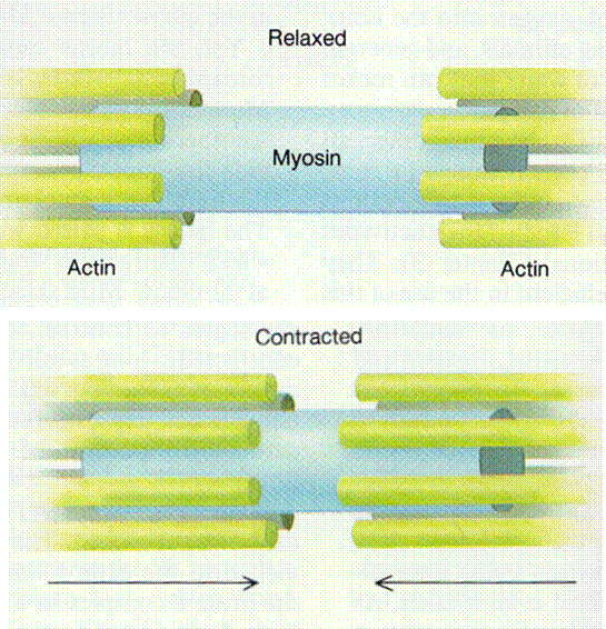
In the relaxed state, regulatory systems prevent actin-myosin interactions. This inhibition is overcome through nervous stimulation, increasing the calcium ion concentration in the myoplasm, which causes a change in the myofilament structure. This enables contraction. Other regulating mechanisms remove calcium ions from the myoplasm to produce the relaxed state.
Membrane Proteins
In both the Sodium (Na), Potassium (K)-ATPase of the plasma membrane and the Calcium ion pump protein (Ca-ATPase) of the sarcoplasmic reticulum membrane
, ATP is split on the cytoplasmic face of the membrane, and some of the energy liberated is used to pump ions in specific directions across the membrane. In the case of the Na, K-ATPase, K is pumped into the cell, and Na is pumped out. Whereas the Ca-ATPase actively pumps calcium into the sarcoplasmic reticulum.
Because of their charge, ions are relatively insolvent in lipids. Lipid membranes have low permeability to most ions. Ionic diffusion across membranes occurs through protein channels. Some channels are highly specific, while others allow passage of all ions below a certain size. Some channels are controlled by the voltage difference across the membrane, and others are regulated by neurotransmitters or other molecules.
Glucose
Glucose has a low solubility in membrane lipids. Glucose enters cells by way of a specific glucose transport protein in the plasma membrane. Monosaccharides enter muscle cells by a facilitated transport process. Monosaccharides compete for the same carrier.
Sugar transport in muscle cells is stimulated by insulin. In the absence of insuline, glucose transport is rate limiting for glucose use. Insulin is a major physiological regulator of muscle glucose metabolism.
Myopathies are disorders of Muscle Function.
References:
Berne, R. M., and Levy, M. N. (1993). Physiology, 3rd Ed.. St. Louis, Missouri: Mosby.
Guyton, A. C. (1991). Textbook of Medical Physiology, 8th ed. Philadelphia: W.B. Saunders Co.
|